Anatomy Label Major Arteries And Veins ~ 32 Veins Of The Body Diagram - Wiring Diagram List. People of all ages benefit from visual learning. This illustration was published in. Figure 47.14 label the major systemic arteries. This is a tutorial on the heart and some of the major vessels that lead to the heart and from the you've got the right brachiocephalic vein and the left brachiocephalic vein. Major arteries, pulse points, and veins.
Labels include cephalic vein, brachial artery/vein, basilic vein, musculoskeletal nerve, ulnar collateral artery note the names of the major veins and arteries involved.(e.g., carotid arteries and jugular veins for the head). We hope this picture major arteries of the body can help you study and research. Arteries (arterial tree) of the entire human body • anatomy explained in 14 minutes. Veins have thinner walls than arteries. The superficial branch is a cutaneous nerve that runs under the brachioradialis muscle and passes through the anatomical snuff box, which is a visible depression formed near the base of the thumb by the tendons.
Jennifer Kersey E-Portfolio Bio211: 2011-04-10 from 1.bp.blogspot.com In fact, many are so tiny only one blood cell can move. Learn anatomy faster and remember everything you learn. Iv = internal jugular vein. We hope this picture major arteries of the body can help you study and research. The superficial branch is a cutaneous nerve that runs under the brachioradialis muscle and passes through the anatomical snuff box, which is a visible depression formed near the base of the thumb by the tendons. These carry oxygenated blood from the heart to the rest of the body. Arterioles further branch into capillaries, the true deliverers of oxygen and nutrients to your cells. The veins arteries and capillaries labeled sticky anatomy wall chart is perfect for reporting findings, consultations, and procedural explanations.
Iv = internal jugular vein.
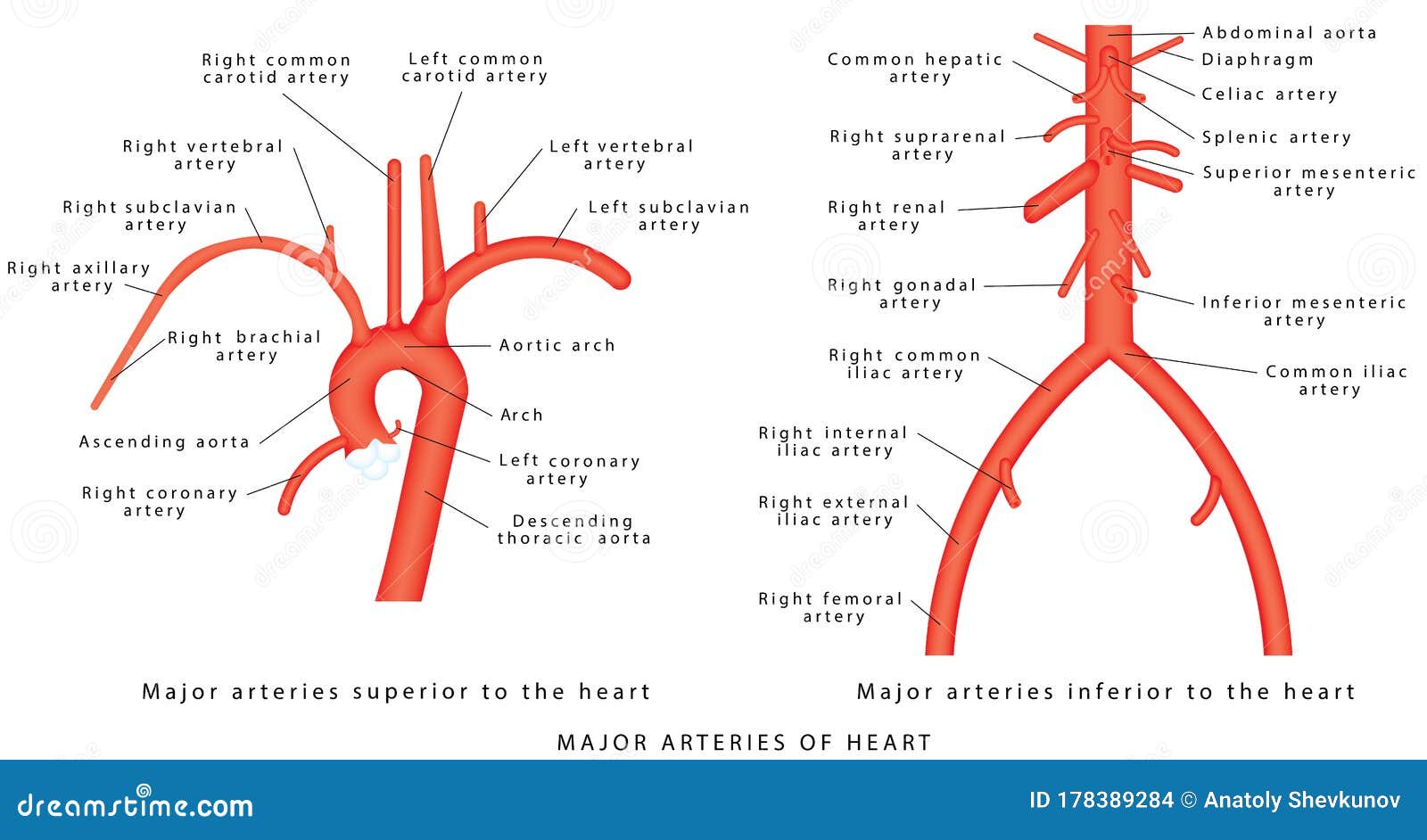
The arteries are strong, muscular, and stretchy, which helps push blood veins: Explore the anatomy of the human cardiovascular system (also known as the circulatory system) with our detailed diagrams and information. The abdominal aorta bifurcates at the level of the fourth lumbar vertebra to form the two common iliac arteries, each of which further branches into the external and the internal iliac artery. In human anatomy, the axillary artery is a large blood vessel that conveys oxygenated blood to the lateral aspect of the thorax, the axilla (armpit) and the upper limb. The major deep veins of the arm are the radial and ulnar veins, which run along the length of their respective bones and merge at the elbow to form the. As compared with those of the arteries, diseases associated with the veins are often very common, curable, and hardly fatal. People of all ages benefit from visual learning. The superficial branch is a cutaneous nerve that runs under the brachioradialis muscle and passes through the anatomical snuff box, which is a visible depression formed near the base of the thumb by the tendons. Laboratory manual for human anatomy & physiology | 2nd edition. Because arteries are moving blood being pumped below are some of the major arteries that are found in the body and the organs and tissues that this has three major branches — the brachiocephalic trunk, the left common carotid artery, and the left. Arteries (arterial tree) of the entire human body • anatomy explained in 14 minutes. Iv = internal jugular vein. Testicular (gonadal) vein (direct tributary on right side;
In human anatomy, the axillary artery is a large blood vessel that conveys oxygenated blood to the lateral aspect of the thorax, the axilla (armpit) and the upper limb. From there, blood passes through major arteries. Major arteries, pulse points, and veins. Veins have thinner walls than arteries. The superficial branch is a cutaneous nerve that runs under the brachioradialis muscle and passes through the anatomical snuff box, which is a visible depression formed near the base of the thumb by the tendons.
artery and vein histology labeled - Google Search | Arteries and veins, Collagen fibers, Arteries from i.pinimg.com This illustration was published in. It will empower your patients and give you the tools to instill confidence and trust. Its origin is at the lateral margin of the first rib, before which it is called the subclavian artery. The superficial branch is a cutaneous nerve that runs under the brachioradialis muscle and passes through the anatomical snuff box, which is a visible depression formed near the base of the thumb by the tendons. We think this is the most useful anatomy picture that you need. You can click the image to magnify if you cannot see clearly. The major glands of the endocrine system, excluding ovaries and testes: For example, the path of blood to and from the kidneys is:
As compared with those of the arteries, diseases associated with the veins are often very common, curable, and hardly fatal.
The duodenum (distal half), jejunum and ileum, cecum and appendix, ascending and transverse colons, and right colic (hepatic) flexure. Together, veins, arteries and nerves define neurovasculature. There are three major types of blood vessels: These carry deoxygenated blood back to the heart, and they increase in size as they get closer to the heart. The left ventricle of the heart pumps oxygenated blood into the aorta. Because arteries are moving blood being pumped below are some of the major arteries that are found in the body and the organs and tissues that this has three major branches — the brachiocephalic trunk, the left common carotid artery, and the left. If you were to lay out all the blood vessels of the body in a line, they would stretch for nearly 60,000 miles. Testicular (gonadal) vein (direct tributary on right side; Thoracic aorta, abdominal aorta, iliac arteries veins: In case of portal hypertension, the superficial veins radiating from the umbilicus (site of portocaval anastomosis) become dilated and tortuous. Laboratory manual for human anatomy & physiology | 2nd edition. Cc = common carotid artery. Supplies the structures of the midgut:
The veins arteries and capillaries labeled sticky anatomy wall chart is perfect for reporting findings, consultations, and procedural explanations. Veins, blood vessels which return blood to the heart, are different in structure and function from the arteries, which carry blood to the circulation. Major arteries, pulse points, and veins. Figure 47.14 label the major systemic arteries. In human anatomy, the axillary artery is a large blood vessel that conveys oxygenated blood to the lateral aspect of the thorax, the axilla (armpit) and the upper limb.
The major arteries stock vector. Illustration of thoracic - 178389284 from thumbs.dreamstime.com Testicular (gonadal) vein (direct tributary on right side; Learn anatomy faster and remember everything you learn. You can click the image to magnify if you cannot see clearly. The external carotid artery supplies the areas of the head and neck external to the cranium. The abdominal aorta bifurcates at the level of the fourth lumbar vertebra to form the two common iliac arteries, each of which further branches into the external and the internal iliac artery. As compared with those of the arteries, diseases associated with the veins are often very common, curable, and hardly fatal. In many instances, the artery and vein that serve the same organ have the same name. Vein located at the side of the neck to collect blood from the brain and parts of the face and neck.
Arterioles further branch into capillaries, the true deliverers of oxygen and nutrients to your cells.
If you were to lay out all the blood vessels of the body in a line, they would stretch for nearly 60,000 miles. Testicular (gonadal) vein (direct tributary on right side; These carry deoxygenated blood back to the heart, and they increase in size as they get closer to the heart. Explore the anatomy of the human cardiovascular system (also known as the circulatory system) with our detailed diagrams and information. Supplies the structures of the midgut: People of all ages benefit from visual learning. Hansen, phd chapter:introduction to the human body page:14. There are three major types of blood vessels: From there, blood passes through major arteries. Major arteries, pulse points, and veins. Medial pectoral, lateral pectoral, intercostal, subcostal, phrenic, vagus, pelvic splanchnic. Anatomy of the nerves, arteries and veins of the arm (upper extremity). This illustration was published in.
Share :
Post a Comment
for "Anatomy Label Major Arteries And Veins ~ 32 Veins Of The Body Diagram - Wiring Diagram List"


Post a Comment for "Anatomy Label Major Arteries And Veins ~ 32 Veins Of The Body Diagram - Wiring Diagram List"